Achilles Spur
Robert H. Sheinberg, D.P.M., F.A.C.F.A.S., D.A.B.F.A.S.
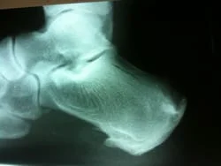
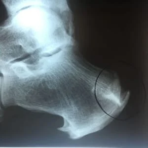
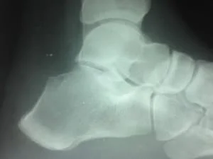
Below is an x-ray demonstrating a lateral view of the heel bone where one can see spurs on the bottom of the heel near the origin of the plantar fascia and at the origin of plantar foot muscles. There is also a spur at the back of the heel where the Achilles attaches. These occur over a long period of time because of excess stress applied to the areas due to tight ligaments and muscular structures.
Discussion:
- Inflammation can be related to a Haglund deformity (postero-superior prominence - normal variant) which causes overlying bursitis.
- The prominence of the posterior superior calcaneal tuberosity contributes to inflammation of the overlying tissues and the Achilles tendon.
- Most often occurs in women and is related to shoe wear with rigid heels or heel counters.
- Patients note posterolateral prominence and tenderness.
MRI:
- Useful to determine if there are distinct degenerative areas within the tendon (Achilles tendinosis), which might require debridement if resection of the Haglund's deformity were indicated.
Non-Operative Treatment:
- Nonoperative treatment consists of heel cord stretching, change in shoe wear, NSAIDs.
-
Raising the heel out of the shoe with a heel insert shifts the
contact against the heel and often relieves symptoms.
Operative Treatment:
- Excision of the Haglund prominence can be effective in chronic cases.
- Excision must be kept proximal to the Achilles insertion.
- Lateral approach is easier but care must be taken to avoid sural nerve.
- The posterior calcaneal tuberosity is removed and the Achilles tendon is debrided and reattached using bone anchors.
- Calcium deposits are removed from the Achilles tendon if they are present.
- Patients are immobilized for six weeks.
These are intraoperative pictures of removal of a painful spur in the back of the heel bone that failed conservative treatment.
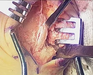
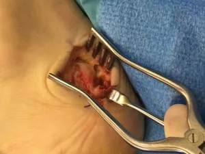
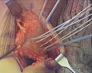
This is an intraoperative pic of a heel spur in the back of the heel prior to resection. It is next to the instrument on the bottom right. The spurs cause pain, as it irritates the Achilles tendon.
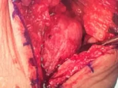
This is the bone cut performed to remove the spur and bone that causes pain.
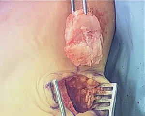
This is a picture of the bone that is removed. Underneath the bone is the void left after removal.
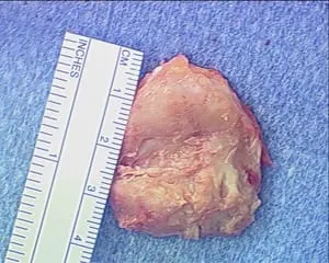
This is a picture of the size of the bone removed. The top portion should have glistening white cartilage. It is yellowish and devoid of cartilage due to constant irritation from the Achilles. The spur is at the bottom 30% portion of the bone.
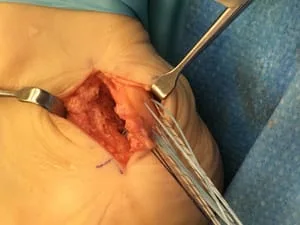
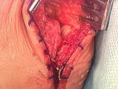
This is a picture of the suture placement in the Achilles tendon to reattach the tendon to bone. There are anchors in the bone that have the suture already attached. The anchors are driven into the bone to allow the Achilles tendon to reattach to the heel. These sutures are then tightened down for an intimate bone to tendon apposition.
Resection of hypertrophic heel spur in the back of the heel that is removed.
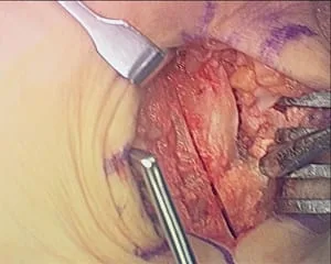
In this pic, the Achilles can be seen on the right being pulled from the back of the heel. The Achilles becomes irritated due to chronic pressure against the heel bone

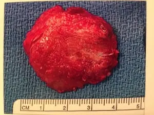
This is the piece of bone removed from the back of the heel.
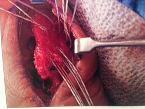
These are the sutures going through the Achilles to reattach to the back of the heel bone.
Pre and PostOp Heel Spur resection with reattached of Achilles.
Pre and postop heel spur resection with reattachment of Achilles.
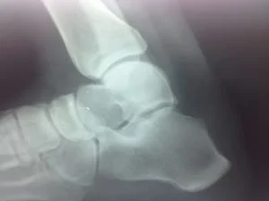
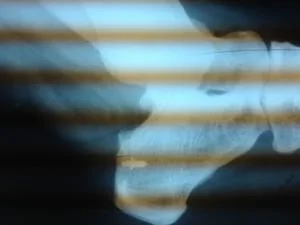
Postop bilateral calcaneal spur resection.
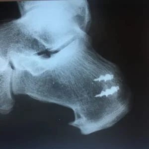
Postop x-ray after spur resection with reattachment of Achilles with the G2 anchor.
Intraop Pics of Posterior Achilles Spur Excision and Reattachment of Achilles Tendon