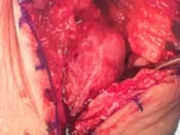
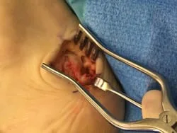
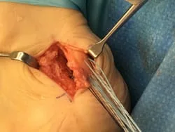
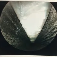
These are intraoperative pictures of removal of a painful spur in the back of the heel bone that failed conservative treatment.
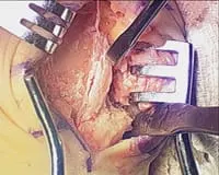
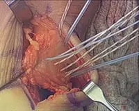
This is an intraoperative pic of a heel spur in the back of the heel prior to resection. It is next to the instrument on the bottom right. he spur causes pain, as it irritates the Achilles tendon.
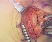
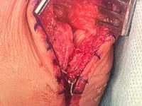
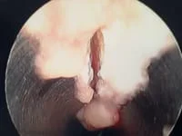
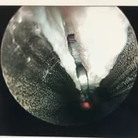
This is the bone cut performed to remove the spur and bone that causes pain.
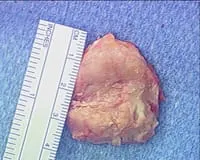
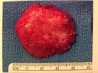
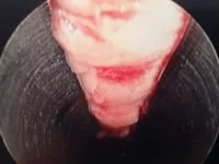
This is a picture of the bone that is removed. Underneath the bone is the void left after removal.
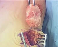
This is a picture of the size of the bone removed. The top portion should have glistening white cartilage. It is yellowish and devoid of cartilage due to constant irritation from the Achilles. The spur is at the bottom 30% portion of the bone.
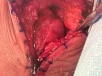
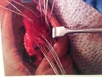
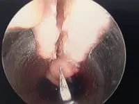
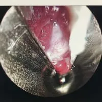
This is a picture of the suture placement in the Achilles tendon to reattach the tendon to bone. There are anchors in the bone that have the suture already attached. The anchors are driven into the bone to allow the Achilles tendon to reattach to the heel. These sutures are then tightened down for an intimate bone to tendon apposition.
Resection of hypertrophic heel spur in the back of the heel that is removed.
In this pic, the Achilles can be seen on the right being pulled from the back of the heel. The Achilles becomes irritated due to chronic pressure against the heel bone
This is the piece of bone removed from the back of the heel.
These are the sutures going though the Achilles to reattach to the back of the heel bone.
Pre and postop heel spur resection with reattachment of Achilles.
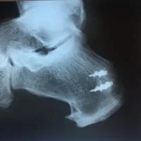
Postop bilateral calcaneal spur resection.
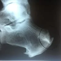
Postop x-ray after spur resection with reattachment of Achilles with G2 anchor.

Intraop Pics of Posterior Achilles Spur Excision and Reattachment of Achilles Tendon
Pictures during an endoscopic plantar fasciotomy or release. The medial band of the fascia is visualized on the left. The two right pics are during the release of the band.
More Intraop Pics of Endoscopic Plantar Fascia Release. The thickened fascia is seen on the left. The Blade is cutting through the fascia in the center pic and confirmation of complete release of that portion of the ligament is noted as we can see the intrinsic muscles after release.
ENDOSCOPIC CALCANEOPLASTY FOR “CALANEO-ACHILLES IMPINGEMENT”
By Robert H. Sheinberg, D.P.M., F.A.C.F.A.S., D.A.B.F..A.S.
What is it?
- Endoscopic (with a miniature camera and small incisions) surgical removal of a bony prominence in the back of the heel.
Causes of this bony prominence and pain:
- Enlarged bone called the calcaneus that presses on the front portion of the Achilles tendon.
- Usually seen in high arched feet.
- Associated with “Haglund deformity” or “pump bump”.
- Injury of heel bone or a fracture causing displacement of the top of the heel bone backwards. This causes irritation of the bone against the Achilles tendon.
- Chronic bursitis (retrocalcaneal) caused by friction between the Achilles tendon and the heel bone.
- Associated with a foot type called a rearfoot varus in which the heel bone has a slight curvature inward.
Signs and symptoms:
- Painful enlarged bump on the back of the heel bone.
- Swelling is often present and associated with a bursitis.
- Always tender to touch the bony prominence.
- Often associated with an overlying callus or hard skin over the area.
- Pain is often present in closed shoes as the heel bone presses against the back of the shoe.
- Pain is often present while moving the foot up (dorsiflexion).
Advantages of an endoscopic surgical procedure:
- Minimally invasive surgery with two incisions approximately 1/6” in length.
- Decreased risk of complications including infection and nerve injury.
- Quick return to shoes.
- Faster return to sports and activities.
- Does not weaken the Achilles tendon postoperatively.
- Decreased postoperative pain.
Imaging used for diagnosis:
- X-rays:
- Often see an enlarged bone in the back of the heel.
- A loss of the soft tissue plane on x-rays indicative of bursitis and swelling.
- A high inclination of the heel bone.
- May see calcification of the Achilles tendon which may or may not be associated with the pain or problem.
- Ultrasound:
- Often used to visualize the retrocalcaneal bursa in thickening of the Achilles tendon.
- Used dynamically to see the impingement of the Achilles tendon against the heel bone.
- MRI:
- Used to identify bone marrow edema that is present in the area of the impingement of the tendon against the heel bone.
- Used to identify any associated Achilles tendon damage that may be present.
- Used to visualize the retrocalcaneal bursa and any associated soft tissue pathology.
- Used to visualize any associated bone deformity that may be present.
Testing:
- Local anesthesia may be injected directly into the bursa between the tendon bones. Relief of pain for a few hours will help distinguish this condition from other associated problems and give us an indication of the benefits of cervical decompression by an “Endoscopic Calcaneoplasty”.
Treatment:
- A thorough history and physical examination is performed.
- Evaluation of any bone or soft tissue abnormality is identified.
- Gait analysis to identify any abnormality that may cause the impingement or aggravate the problem line.
- X-rays, MRIs and ultrasounds are often performed to help complete the examination.
- Open surgical procedures can be performed to decompress the area.
- “Endoscopic calcaneoplasty to remove the bony prominence and decompress the Achilles tendon from pressuring the heel bone.
How is it performed:
- Outpatient surgical procedure under a local, twilight or general anesthetic. This is usually the patient’s choice as to the type of anesthesia that they desire to use.
- Two small incisions approximately 1/6” in length are made on each side of the Achilles tendon.
- Specialized instruments including a small camera and surgical burs are used to visualize and remove the bony prominence of the heel bone against the Achilles tendon. If the Achilles tendon has some damage, this can be cleaned up as well.
- One stitch is used to close each incision.
- Surgical boot for 3 to 28 days may be necessary depending on the amount of bone that is resected.
- Early range of motion begins the next day after surgery to prevent adhesions.
- Physical therapy may be started in three days to restore range of motion and strength and to decrease swelling.
- Swimming may be performed in seven days.
- Running may begin on the treadmill in 7 to 28 days.
- Most sports can be resumed in 2 to 6 weeks.
Prognosis:
- Excellent in almost all cases with minimal risk.
Please view more Calcaneoplasty Videos on our youtube page