Our Surgeons are trained in the latest, most advanced techniques: Minimally Invasive Achilles Tendon Repair

Below are intra-operative pictures of an Achilles tendon repair. The image to the left is a tendon that has been prepared to be reattached with sutures in place and tendon cleaned of diseased tendon. The picture to the right is the tendon re-approximated with sutures tied.


Traumatic Achilles Tendon Disorders
By Robert H. Sheinberg, D.P.M., F.A.C.F.A.S., D.A.B.F.A.S.
The Achilles tendon is the strongest and largest tendon in the human body. It is formed by the gastrocnemius and soleus muscles (calf), and inserts into a broad fashion to the back of the heel (calcaneus). The primary function of this muscle-tendon unit is to provide powerful propulsion in walking or running or powerful jumping.
The blood supply to the mid-portion of the tendon is poor, making the tendon susceptible to injury. Injuries may be in the form of inflammation to the tendon covering (paratenonitis), a disease of the tendon (tendinosis), spurring at the heel (insertional spurs with tendinitis) and partial or complete ruptures of the Achilles tendon.
Susceptibility to the injury in sports or at work can often be predicted. A tendon will become inflamed (tendinitis) if an excessive load is placed upon it. This may occur in a worker who is constantly going up and down on their toes to reach for something when they are not accustomed to it or climbing up and down a ladder too frequently in a short period of time. It may also occur if the worker has to push heavy items, walks excessively up an incline or jumps up and down too frequently in a short period of time. We often see it in weekend athletes or joggers who try to get into shape too quickly and do not stretch. Tightness in the Achilles tendon makes one more susceptible to these injuries and poor shoe gear may contribute to this as well.
Ruptures of the tendon, complete or partial, may occur when a worker or athlete suddenly moves the foot up (dorsiflexion) when slipping on a stair or stepping quickly. It may also occur if the front of the foot moves violently up, putting extreme stress on the tendon. The tendon tear tears just as a rope would if the ends of the rope are pulled apart. Tendinitis may weaken the tendon, making it more susceptible to rupture. Tendinitis may often precede weeks or months before the tear.
Stiffness and pain in the Achilles tendon after getting out of bed in the morning or after sitting for a short period of time is the most common complaint. As the person walks the pain and stiffness will often diminish as the tendon stretches and the blood supply to the area improves. As the condition worsens, the stiffness and pain last longer in the morning and burning and discomfort develop with excessive weight bearing. Occasionally, swelling or a crackling sensation may be felt in the back of the Achilles tendon or heel area. If a partial tear has developed, a small tender nodule may be felt on the tendon.
TREATMENT: When tendinitis develops, treatment should be aimed at reducing the inflammation and changing the work status or activity of the person to avoid chronic conditions and tendon ruptures. Job and activity descriptions that include more sitting, less pushing, less reaching and less walking will often allow the tendon to rest until the inflammation subsides. Heel lifts, non-steroidal anti-inflammatories, ice and physical therapy may be necessary. Occasionally day and night splints are used. Avoiding walking barefoot is essential. Once the acute inflammation subsides, stretching will often stimulate flexibility, decreasing the likelihood of recurrence. Depending on the severity of the condition, it may last one to eight weeks. If symptoms do not reduce, immobilization in a Cam Walker may be necessary. Ruptures of the Achilles tendon should be treated surgically on most occasions. Cleaning the ends of the tendon and re-approximating the edges is the treatment of choice. Postoperative immobilization is necessary to avoid stress on the operative area. The immobilization process is 1-3 weeks and is followed by physical therapy to achieve 100% return to activity.
Achilles tendon ruptures are the most common tendon ruptures of the lower extremity. They can occur at any age, but are most common in the third to fifth decade. There is a significant male preponderance. The classic description is the "weekend warrior" athlete.
Anatomy
The Achilles tendon is the common tendon of the gastrocnemius and soleus muscles and provides their attachment to the calcaneus. The soleus muscle arises from the posterior tibia while the gastrocnemius arises from the posterior distal femur. This allows the gastrocnemius to be effective with an extended knee and the soleus to be more effective with a flexed knee.
The tendons from both muscles coalesce just distal to the musculotendinous junction to form the Achilles tendon. The tendon has a relative avascular portion 2-6 cm above the insertion. The tendon also rotates approximately 90 degrees during its course, with the gastrocnemius fibers being more lateral. The tendon inserts upon the posterior calcaneus primarily along the posterior tuberosity with slightly more medial than lateral extension.
Pathogenesis
A relatively hypovascular area exists approximately 2-6 cm above the insertion into the calcaneus. This hypovascularity has been implicated in disorders of the tendon. Age-dependent changes in collagen cross-linking result in increased stiffness and loss of viscoelasticity, which may predispose the tendon to rupture. Mechanisms associated with ruptures include suddenly forced dorsiflexion of the ankle (eccentric contraction of the gastrocnemius and soleus), pushing off with the weight-bearing forefoot while extending the knee, and laceration or a direct blow to the contracted tendon.
Classification
Achilles tendon ruptures are partial or complete. Ruptures can also be divided into acute traumatic ruptures, chronic ruptures, or chronic attritional ruptures. However, ruptures are often due to a combination of age-related attrition and an acute traumatic incident.
Patient History and Physical Findings
The patient with Achilles tendon rupture presents with pain in the area of the Achilles tendon. The pain of an acute rupture is often described as an intense burning sensation or sharp stabbing pain. Patients may hear an audible pop after an eccentric muscle contraction or be pushing off; they usually describe a feeling of being kicked, hit, or shot in the heel. A small percentage of patients will have prodromal symptoms. In the presence of a complete tear, patients will experience significant ankle plantar flexion weakness. However, many patients continue to be able to actively plantarflex the ankle using accessory muscles. This may confound some examiners and result in a missed diagnosis.
Physical findings include a visible soft-tissue depression in the posterior ankle on observation. The tendon defect can often be palpated along the posterior leg and ankle. Patients may be unable to walk or walk only with a limp secondary to weak or absent push-off. The absence of active plantarflexion is often expected, but many patients effectively recruit other muscles to plantarflex against manual testing. However, they are rarely able to perform a single leg heel raise. With the patient in the prone position and the knees flexed, the Thompson squeeze test is executed by squeezing the calf muscle and observing the presence or absence of resultant ankle plantarflexion and comparing with the contralateral side. Another helpful test is to observe the resting position of the ankle compared to the unaffected side with the patient prone and the knees flexed to 90 degrees.
Imaging and Diagnostic Studies
Radiographs are rarely diagnostic. They may be warranted in cases of extremely distal ruptures when avulsion of part of the calcaneus needs to be ruled out. Ultrasound and MRI can accurately demonstrate ruptures but are rarely necessary with classic clinical findings. These studies may be helpful when the diagnosis is unclear.
Treatment
Treatment for acute Achilles tendon ruptures can be operative or non-operative and much controversy exists. Historically, the pendulum swung towards operative treatment (especially of younger, healthier patients) because of the much lower reported re-rupture rate (2% for surgical and 11-30% for non-surgical), accepting the trade-off of potential wound complications.
Non-operative Treatment
Conservative treatment varies and classically involved casting in a long leg cast with the knee flexed and ankle in equinus (2-3 weeks), then short leg casting (8 weeks). Nonweightbearing was typically recommended initially (the first 6 weeks).
More recent approaches include functional bracing with immediate weight-bearing. These more aggressive protocols describe immediate full weight-bearing in a functional brace or pre-fabricated boot. Patients are started with the ankle in up to 45 degrees of plantarflexion, which is gradually decreased to neutral over 6 to 12 weeks. They often perform active plantarflexion exercises with restricted dorsiflexion during that time, then graduate to more aggressive strengthening protocols.
Operative Treatment
At South Florida Institute of Sports Medicine, the Foot and Ankle Surgeons prefer open operative repair. Depending on the existing tendon condition and timing of repair, cast immobilization varies from 2-6 weeks. Weight-bearing begins at 4-6 weeks and intensive physical therapy follows with a Cam Walker transitioning back to shoe gear.
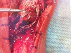
Intraop pic of Achilles ruptures prior to repair. There is a 1-cm gap between the tendon ends. The white tendon on the left is called the plantaris. Due to this tendon being intact, the Achilles didn't retract much after the rupture.
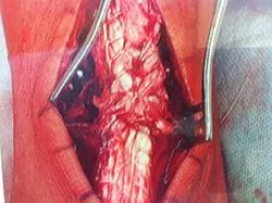
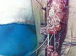
Pictured below is an image of a torn Achilles tendon. The ends are frayed because this is a result of a chronic degenerative process similar to when a rope starts to wear out and then finally gives out.
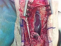
This is the same tendon after surgical repair.
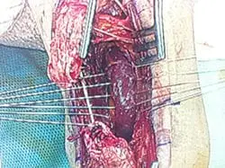
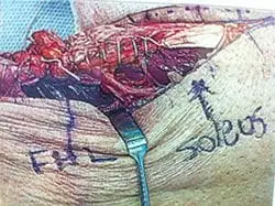
Series of pics during Achilles tendon rupture repair with posterior fasciotomy and FHL muscle transposition to increase vascularity to the repair site (below). The FHL Muscle transposition is a novel approach, devised by Dr. Robert Sheinberg over 15 years ago, that the practice continues to use to have excellent results and added vascularization to the Achilles tear.

Torn Achilles Tendon

Identifying the posterior fascia that separates the Achilles from the
FHL muscle and compartment.


FHL muscle is sutured across to bring better blood flow to the repair
site.


Krackow suture of both Achilles rupture stumps


Rupture is repaired and the FHL muscle can be seen as one with the
Achilles to allow increased blood flow for healing.
Series of pics during Achilles tendon rupture repair with posterior fasciotomy and FHL muscle transposition to increase vascularity to the repair site (below).
Intraop picture of Achilles tendon rupture. We have made a posterior fasciotomy to visualize the FDL muscle. We have 4 sutures in the center of the pic that are in the muscle belly. We tie these sutures on the pic of the tendon after the repair. This gives a better blood supply to the repair site.
Pic of Rupture. The white tendon to the left is called the plantaris tendon. It doesn't give any tension to the Achilles or repair.

Windowing and Fasciotomy to Expose the FHL muscle


Pics of the Sutures through the muscle to transpose to the Repair site of
the Achilles tear


Krackow suture technique to lock the 2 ends of the repair site to
decrease tension to the repair.


Repaired Achilles tendon with the sutures on the left and right holding
the FHL muscle to transpose to the repair.


Pics of the complete repair and FHL muscle transposition to increase
vascularity and healing to the repair


Series of pics during Achilles tendon rupture repair with posterior
fasciotomy and FHL muscle transposition to increase vascularity to the
repair site (below).
The rupture ends are at the top and bottom of the pic.

The window is made in the fascia between the Achilles and FHL muscle (the muscle that moves the big toe down). We pass suture into the muscle and this can be seen in the fingers on the left and right of the pic.

We have sutured the ends of the Achilles tear and these will be sutured together to repair the tear.


The tendon is tied together and the suture placed in the FHL muscle is tied around the tendon to transpose the muscle to the tendon repair to increase vascularization to the tendon and promote better healing.


The rupture ends are at the top and bottom of the pic. This is the fascia between the Achilles and the deep posterior compartment.


Below is a pic of the window we make in the fascia to get to the FHL muscle.

We weave absorbable suture through the muscle so after the repair we attach the muscle to the front side of the Achilles to increase vascularity to the repair.
Below are the pics of the Krackow suture technique we use to repair the proximal and distal repair stumps together. We interweave a non-absorbable suture through each rupture end and then tie them together to reapproximate the tendon.


We have repaired the Achilles and are preparing to transpose the FHL muscle to the repair site to increase vascularity. The four sets of the white suture can be seen on the sides of the Achilles.


Pic after complete repair with muscle transposition

The instrument is identifying the FHL muscle as it is transposed to the repair site to increase vascularity.

Another case of a torn Achilles tendon as a result of chronic degeneration of the tendon fibers.

In the picture above you can see the muscle belly of the Flexor Hallucis Longus which can provide better blood supply to the area of the tendon being repaired as it has been shown, histologically to have decreased blood flow compared to the rest of the Achilles tendon.
The muscle is prepared with multiple sutures with free ends to then lay the repaired tendon over, then securing the tendon with the suture.
This is the tendon being repaired with suture using a technique called a Krachow Stitch.
This is an image of the repaired tendon, now in one piece overlying the Flexor Hallucis Longus (FHL) muscle belly with the previously placed sutures securing the tendon against the muscle.
Intraop picture of Achilles tendon rupture. We have made a posterior fasciotomy to visualize the FDL muscle. We have 4 sutures in the center of the pic that is in the muscle belly. We tie these sutures on the pic of the tendon after the repair. This gives a better blood supply to the repair site.
Achilles tear


Pic of Fascia between Achilles and FHL muscle prior to fasciotomy to expose the FHL Muscle
Posterior Fasciotomy (Window) with FHL muscle exposed and the sutures seen at the right and left that will transpose the muscle to the repair.


Krackow suture technique on both sides of the Achilles tendon that locks the tendon on both sides of the Achilles to decrease tension at the repair site.

Repair of the Achilles. The sutures on the sides are attached to the FHL muscle prior to being tightened to transpose the muscle.
From the side, the FHL muscle can be seen transposed to the repair to increase vascularity and healing to the tendon.


Intraop picture of Achilles tendon rupture. We have made a posterior fasciotomy to visualize the FDL muscle. We have 4 sutures in the center of the pic that is in the muscle belly. We tie these sutures on the pic of the tendon after the repair. This gives a better blood supply to the repair site.

This is a close-up view of the repair and the muscle adhered to the front of the tendon.

View of the repair from the top.
Repair of traumatic Achilles tendon rupture with Krackow suture technique and FHL muscle transposition.
View of the rupture


View of the posterior fasciotomy and exposed FHL muscle to increase vascularity to the repair.


We have placed absorbable suture through the muscle belly to later transpose to the front of the repair.

Achilles rupture has been repaired with a Krackow technique and the absorbable suture can be visualized from each side of the Achilles prior to suturing.

Repair with transposition of the FHL muscle to increase vascularity to the repair site.
(The instrument to the left of the Achilles is identifying the FHL muscle.)


Series of pics of Achilles rupture with transposition of FHL muscle to improve vascularity to the repair.
Achilles Rupture

Posterior fasciotomy to expose the FHL muscle for transposition.

Sutures in the FHL muscle belly for transposition.

Achilles is repaired and the sutures on each side of the tendon are to transpose the FHL muscle to the repair site.

Final repair with transposition. The instrument to the left is showing the FHL muscle belly transposed to the repair.

Achilles ruptures intraoperatively before and after repair.


Intraop pic of Achilles ruptures prior to repair. There is a 1-cm gap between the tendon ends. The white tendon on the left is called the plantaris. Due to this tendon being intact, the Achilles did not retract much after