Open Fasciotomy for Acute Compartment Syndrome
Pictured below is an opening of the facial muscle compartments of the left leg. This procedure is performed to release pressure that has built up in the muscle from activity in which the pressure does not go down after a certain amount of time. This increased, sustained pressure within the compartments is very painful and puts at risk loss of blood supply to the muscles, and in severe cases can cause the death of the muscle tissue.
Below is a picture of retention sutures placed over the fasciitomy site in order to allow the muscle compartment to decompress but also does not allow the skin to retract and make closure of the large inscision difficult to perform at a later date.

This is primary closure of the incision site once the compartment has been adequately decompressed,


Compartment Syndrome
The following is the fascial covering over a muscle compartment. The condition occurs when the pressure within this compartment builds up after an injury or can also occur after exercise from the inability of the muscle to decompress naturally. This leads to pain that is unrelenting and could lead to decrease blood flow and oxygen to muscle tissue causing the death of that tissue.

The picture below demonstrates the fascia overlying the tissue being lifted by the forces at the top of the picture.

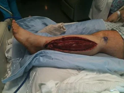
Below is demonstrates incision into the compartment exposing the muscle tissue that is a deeper red color where the retractor is to the left of the picture. The opening of this facial tissue is vital in releasing pressure in the compartment after injury or from exercise. It also important that this release is extensive as so the fascial tissue does not heal closed allowing build of pressure again.

Achilles Tendon Rupture
Most Achilles tendon tears occur from chronic degeneration of the tendon in conjunction with the improper conditioning of the tendon to perform certain activities such as jumping and sudden changes in direction. This is why this type of injury occurs in what is considered the "Weekend Warrior". This person usually does not train and condition the tendon to handle intense physical activity then goes and plays a sport over the weekend and the tendon gives out. In the picture below one can see the frayed ends of the torn Achilles tendon.

The image below is the tendon after repair with a complex suture technique called the Krakow stitch. This is a very strong repair and prognosis after repair with physical therapy is very good.

Series of Pics During Achilles Tendon Rupture Repair with Posterior Fasciotomy and FHL Muscle Transposition to Increase Vascularity to the Repair Site (Below)
The Rupture ends are at the top and bottom of the Pic. This is the fascia between the Achilles and the Deep Posterior Compartment.

Below is a pic of the window we make in the fascia to get to the FHL muscle

We weave absorbable suture through the muscle so after the repair, we attach the muscle to the front side of the Achilles to increase vascularity to the repair.

Below are the pics of the Krakow suture technique we use to repair the proximal and distal repair stumps together. We interweave a non-absorbable suture through each rupture end and then tie them together to reapproximate the tendon.


We have repaired the Achilles and are preparing to transpose the FHL muscle to the repair site to increase vascularity. The four sets of the white suture can be seen on the sides of the Achilles.

