ARTHRITIS OF THE ANKLE
By: Robert H. Sheinberg, D.P.M., D.A.B.F.A.S., F.A.C.F.A.S.
Causes:
- Trauma to the ankle that could have occurred in childhood and progressed.
- Ankle fracture into the ankle joint.
- Frequent ankle sprains creating a chronically unstable ankle.
- Bone infections (uncommon).
- Synovial tissue disease (rheumatoid arthritis, psoriatic arthritis).
- Gouty arthropathy.
- Diabetes (Charcot neuro arthropathy).
- Tumors that have invaded the joint.
- Congenital deformities of bone to the ankle joint.
- Avascular necrosis (dead bone).
Signs and Symptoms:
- Slow progressive loss of motion of the ankle joint.
- Stiffness and soreness, especially getting out of bed in the morning to begin to ambulate. In the short term the stiffness may go away very quickly but as the disease progresses the stiffness will last longer. Also associated with stiffness after getting out of a car or after sitting in a chair to get up and walk.
- With progression a chronic ache in the ankle joint.
- Painful with motion, especially up and down.
- Pain with ambulation and difficulty running.
- Diffuse swelling to the ankle joint.
- In women, may have difficulty wearing any flat shoes.
X-rays:
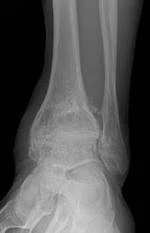
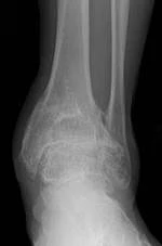
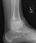
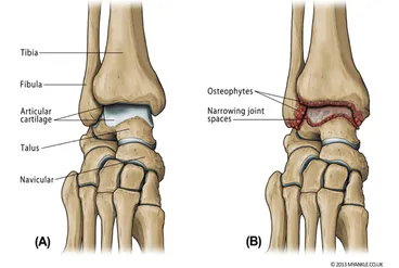
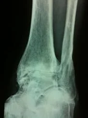
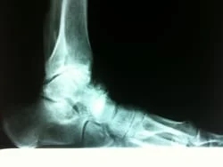
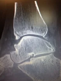
- In mild cases bone spurring may develop around the ankle joint. A small degree of joint space narrowing may also be present. As the arthritis progresses there is a further loss of joint space. The joint surface appears to be white and diffuse bone spurring develops around the joint region.
Treatment:
- Mild cases: Braces - ankle, foot orthoses, (AFO’s) may help to decrease motion in the ankle joint, lessening the pain.
- Moderate cases: When conservative case has not helped, arthroscopic surgery to remove the abnormal bone, soft tissue and cartilage may be of benefit. If the disease process is advanced, only temporary benefit may be achieved.
- Severe cases: When the arthritis has advanced and has been unresponsive to conservative care and/or arthroscopy, fusion of the ankle joint is the gold standard. During the fusion the cartilage and the joint surface is completely removed. The bones are then put together and held in place with screws. This procedure can be performed arthroscopically if there is minimal deformity to the foot and ankle. If there is severe deformity the procedure may be performed open. Long-term outcome is excellent following the procedure with regards to eliminating the pain. Most patients can return to walking without discomfort.
- Some cases of severe arthritis may be candidates for Total Ankle Replacement surgery. This is a technique where the ankle joint is replaced by a prosthetic (artificial) ankle.
The 2 following videos from our YOUTUBE site demostrating the motion of the foot that is possible after an arthroscopic ankle fusion
https://youtu.be/gF58YKmNzKg?list=PLVGxw0HDwtHD2TjHMXgFIEQVTOKdTxGTs
https://youtu.be/d8kBEAXIa7Y?list=PLVGxw0HDwtHD2TjHMXgFIEQVTOKdTxGTs
Our YOUTUBE video below is the gait of a patient 12 weeks after Arthroscopic Ankle Fusion
https://youtu.be/d8kBEAXIa7Y?list=PLVGxw0HDwtHD2TjHMXgFIEQVTOKdTxGTs
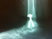
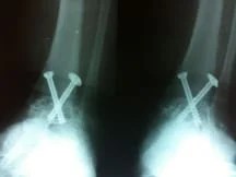
Below are images of post-surgical correction of the ankle arthritis with arthroscopic ankle arthrodesis (fusion) using internal scew fixation
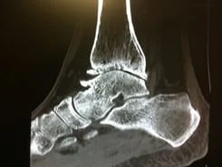
Below is another example of severe ankle arthritis on an x-ray and a sagittal plane CT scan of the ankle that shows more detail of the joint space narrowing, spur formation, subchondral bone cyst in the tibia.
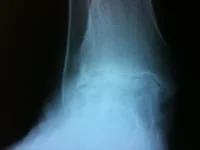
These are pictures of a pilon fracture that was performed by a different surgeon. The anterior tibial plate was placed too distal and invaded the patient's joint. These injuries often cause post-traumatic arthritis regardless of the fixation. The plate was removed and the patient had some relief. Approximately 1 year later, the patient underwent a successful arthroscopic fusion.
S/P Pilon with anterior plate too distal
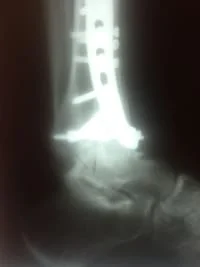
After removal of the plate prior to arthroscopic fusion.
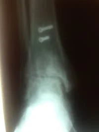
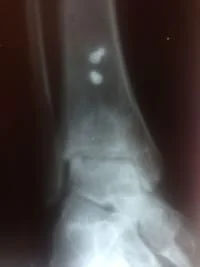
After arthroscopic fusion.
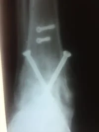
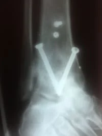
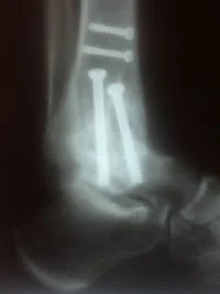
Ankle fusion with fibular onlay graft after severe post traumatic arthritis post pilon fracture.
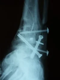
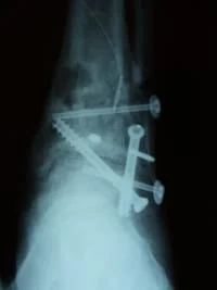
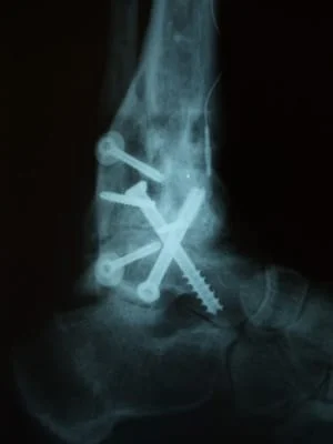
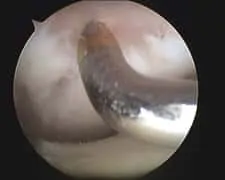
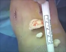
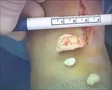
A series of intraop ankle arthroscopy pics of synovial chondromatosis with OCD talus and tibia and microfracture.
Pic of one of the nodules inside the ankle joint.

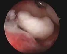
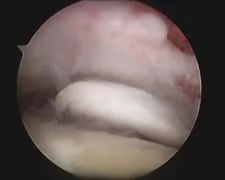
Pic of an OCD in the talar dome due to pressure from the nodule.
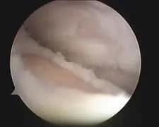
Pic after microfracture and debridement of above OCD.
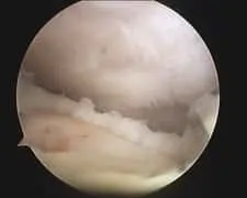
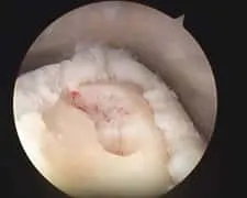
Pic during microfracture of tibial OCD.
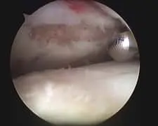
Pics of nodules removed during scope. A separate incision had to be made to remove the nodules due to the size.
Tibial Talar Spurring
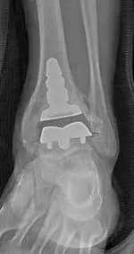
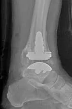
Pre, intraop and postop X-rays status post total ankle replacement for ankle arthritis.
