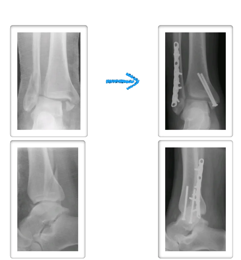
Both the lateral and medial malleolus with fractures with the lateral malleolar fracture classified as a Weber B (at the level of the ankle joint) and the medial malleolar fracture almost transverse (Left x-ray). This is indicative of a Supination External Rotation (SER IV) injury. The fractures are repaired using open reduction with internal fixation (ORIF) technique and fixated with screws and a surgical fractue plate located at the fibular (Right x-ray).

A bimalleolar fracture is a fracture of the ankle that involves the lateral malleolus and the medial malleolus. Studies have shown that bimalleolar fractures are more common in women, people over 60 years of age, and patients with existing comorbidities. Surgical treatment will often be required, usually an Open Reduction Internal Fixation (ORIF). This involves the surgical reduction or realignment of the fracture followed by the implementation of hardware to aid in the healing of the fracture. Usually a plate and screws will be used on the fibular fracture and screws, screws and pins, pins or tension band will most commonly be used on the medial malleolus fracture. A bimalleolar "equivalent" fracture is a fracture of the fibula with rupture of the superficial and deep portion of the deltoid ligaments leaving the medial malleolus intact. Surgical management is common due to the instability of the fracture and displacement of the talus laterally.
Pre and Post Op X-rays of Bimalleolar Fracture ORIF
The x-ray images below demonstrate another case of a bimalleolar ankle fracture in both and oblique view and anterior - posterior view. Open reduction and internal fixation with screws for the medial malleolar fracture and screws and plates for the lateral mallolar fracture. Note the even joint spacing across the ankle mortise after correction that is not evenly spaced in the pre-surgical picture.


Pre and Postop Bimalleolar Fracture

Pre and Postop Bimalleolar Fracture


Cotton Test
This is a fibular fracture that had Deltoid rupture that causes the increased space at the inside (medial) ankle. The joint should be congruous and symmertical. We know the deltoid ligament has been torn due to the medial joint space widening in the first X-ray. This is called a bimalleolar equivalent fracture. After repair of the fibula, an instrument is placed around the fibula and there is traction applied laterally to check the integrity of the syndesmosis. If there is widening of the syndesmosis with lateral pull of the fibula, the test is positive for syndesmotic rupture and will necessitate stabilization of the syndesmosis.


BiMalleolar Equivalent Fracture With Displaced Fibula Fracture and Deltoid Rupture treated with ORIF Fibula with 2 Arthrex Syndesmotic Tightropes.


