CAUSES OF INTOE (PIGEON TOE)
By: Robert H. Sheinberg, D.P.M., D.A.B.F.A.S.,
F.A.C.F.A.S.

A. FEMORAL ANTEVERSION
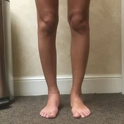
Femoral anteversion is the most common cause of an intoe gait in children greater than 3. It is caused by a twisting of the thigh bone. The thigh bone (femur) has an internal twist when comparing the lower to the upper portion of the bone. This twist is greatest at birth. There is a gentle steady untwisting of the bone that takes place until about age 12. From the ages of 12-16 the change is more subtle as it reaches an adult value.
CAUSE:
- An excess is a developmental abnormality.
- Due to intrauterine position.
- Genetic predisposition.
- Tight hip muscles and hip ligaments contribute to the problem.
CLINICAL EXAMINATION:
- Determining femoral anteversion is usually based on a thorough clinical examination. An examination of the hip range of motion usually reveals that there is a greater internal than external rotation when measuring motion with the hip extended.
- In rare cases x-rays and CT scans may be needed, especially if there is asymmetry of motion between the right and left hip. This may indicate hip dysplasia.
SIGNS AND SYMPTOMS:
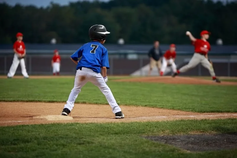
- Intoe gait, more noticeable when a child begins school.
- When standing in the patient’s normal posture, the patella (kneecaps) appear to point in or squint towards each other.
- While observing the patient walk, the knee and patella (kneecap) appear to be turned in. May worsen at the end of the day when the child fatigues.
- Child usually likes to sit in a “w” position.
- Running appears to be awkward.
- If associated with internal tibial torsion (internal position of the lower leg bone on the upper leg bone) a child may trip and fall when running.
- If associated with external tibial torsion (lower leg bone excessive rotated outwardly compared to the upper leg bone) may cause significant malalignment syndrome of the knee.
- More common in girls.
- Usually symmetrical between the right and left sides.
TREATMENT:
The angle of the lower to the upper leg bone steadily diminishes until age 12. The condition usually resolves by that time.
- Observation of the change in position of a child’s knee and foot progression may be all that may be necessary.
- Exercises to stimulate flexibility of the hip towards external rotation.
- Activities, especially ballet for girls and rollerblading for boys and girls may help to improve hip flexibility towards external rotation.
- If asymptomatic no treatment is needed.
- If a condition is excessive (inner rotation greater than 80 degrees and lateral rotation less than 10 degrees) and there is associated tripping or severe cosmetic concern, surgery could be considered after the age of 10. The delay is necessary because of the possible spontaneous resolution of the problem up until the age of 16.
- If associated with external tibial torsion (excessive motion of the lower leg on the upper leg to the outside) and significant knee problems develop, surgery may also be needed to realign the extremity.
PROGNOSIS:
There is a natural decrease in the intoe gait associated with femoral anteversion that occurs in 99% of the cases. The patient should be followed and reassurance should be given to the parents that these cases will almost always resolve in time. It is important to ascertain whether or not the child has internal tibial torsion or external tibial torsion associated with femoral anteversion. This may change the prognosis. Femoral anteversion by itself typically does not lead to arthritic changes in the hips or knees and should not be treated surgically unless significant symptoms warrant it.
B. TIBIAL TORSION
It is a rotation of the lower leg bone (tibia) excessively inwards relative to the upper leg bone (femur). It may also be due to an internal twist of the lower portion of the lower leg bone (tibia) relative to the upper portion of the lower leg bone (tibia). Usually noticed between the ages of 2 and 4. It is the most common cause of intoe in the 2 to 4-year-old age group and the condition usually resolves by age 8.
CAUSE:
- Intrauterine position.
- Excessive medial ligamentous and muscular tightness around the knee region.
- Sitting and sleeping postures may perpetuate the problems but generally do not cause them.
SIGNS AND SYMPTOMS:
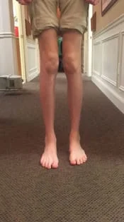
- When viewing the child standing, the foot and lower leg appear to be rotated internally. If there is an isolated problem, the kneecaps appear to be straight, thus distinguishing this condition from femoral anteversion in which the kneecaps are pointed in.
- When walking or running the feet excessively turn in occasionally causing tripping and falling. During running the kneecaps continue to stay straight.
- At the end of the day when fatigue sets in the intoe appears to be worse. May be asymmetrical (one side worse than the other).
- May appear to be bowlegged (because the musculature in the calf is rotated towards the outside of the lower leg).
- Could be associated with metatarsus adductus in an infant.
CLINICAL EXAMINATION:
Examining the child with the patient sitting, standing and walking is important. When the child is sitting the rotation of the lower leg bone is measured against the upper leg bone. When the rotation of the lower leg bone is excessively in with very little rotation out, this is indicative of internal tibial torsion. Observation of the kneecaps is important in helping to rule out femoral anteversion or to determine if there is a component of a hip problem associated with it.
TREATMENT:
- Observation of gait and reassurance to the parents that the condition usually resolves by age 8 is provided.
- Abnormal sitting and sleeping postures which tend to perpetuate the deformity need to be changed.
- A child that is tripping and falling or has a posture that is excessively turning in will benefit from a cast that goes above the knee. During the cast application the lower leg bone is gently rotated externally relative to the upper leg bone. This helps to stretch the ligamentous and musculotendinous structures around the knee. Cast may be utilized for 2-6 weeks. This will rapidly help the normal physiologic unwinding process of the lower leg bone relative to the upper leg bone.
- Night splinting is utilized following cast removal to maintain the correction.
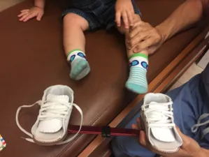
- Counter rotational splints and a Denis Browne bar may also be helpful. They may not help as much in the unwinding process as much as they will help prevent abnormal sleeping postures that could perpetuate the deformity.
- Orthotics and gait plates may also be utilized to help to move the foot towards external rotation during gait. They are also beneficial in preventing abnormal compensation, which could put stress on the arch.
- It is rare that an operation is performed for this condition. If the deformity is excessive causing tripping and falling and there is a significant cosmetic concern, surgery on the lower portion of the lower leg can be performed to externally rotate the foot.
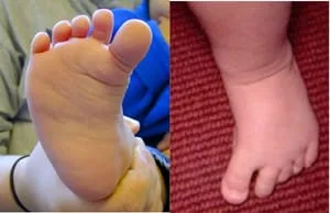
- A deformity of the front part of the foot in which all of the metatarsals are positioned to the inside of the foot.
- Intrauterine position.
- Hereditary predisposition.
- Hyperactive muscle (tibialis anterior, tibialis posterior muscle or abductor hallucis).
- Abnormal bone or joint structure.
- The front half of the foot is turned in relative to the back half of the foot.
- Foot may turn up and in, causing a crease of skin on the inner arch.
- Foot is shaped like a “C” when viewing it from the sole.
- May be associated with rotational problems of the lower leg (internal tibial torsion).
- Excessive space between the first and second toes.
- Prominent bone (fifth metatarsal) on the outside of the foot.
- May be associated with the curvature to the heel bone (calcaneal varus).
- Juvenile bunion and hammertoe deformities.
- Lateral drifting of all the digits as the tendons on the top of the foot pull the toes to the outside.
- Stress fractures to the outside bones in teenagers and adults.
- Arthritis in adults due to the abnormal joint positions.
- Identify the level of the problem (foot, ankle or leg) and the foot’s flexibility.
- Identify any muscle hyperactivity.
- Corrective shoes and occasionally night splinting.
- Casting of the foot and leg to straighten the position of the foot and in some cases the lower leg.
C. METATARSUS ADDUCTUS
CAUSE:
SIGNS AND SYMPTOMS:
ASSOCIATED PROBLEMS:
TREATMENT:
Prognosis is excellent if the condition is treated early.
D. HYPERACTIVE ABDUCTOR HALLUCIS
The abductor hallucis is a muscle that originates on the inner aspect of the
heel bone and forms a tendon that attaches to the inner aspect of the big
toe. Hyperactivity of this muscle causes the big toe to point towards
the inside of the body. This gives it an appearance of having a
metatarsus adductus deformity. These deformities are recognized a few
months after birth and may become more apparent when the child starts to
weightbear. With weightbearing the big toe starts to drift toward the
inside of the foot, causing the appearance of an intoe gait. It is primarily
caused by a muscle imbalance. In most cases, over time the antagonists
to this muscle start to become active, stabilizing the big toe in a more
rectus position. If there is an imbalance of strength between the
inner and outer muscle the big toe may stay in the internal position.
If so, x-rays should be taken to rule out any underlying bone deformity that
could be associated with this problem.
This is a flexible deformity which rarely causes any problems with the child, especially when they are wearing closed shoes. A closed shoe keeps the toe in a more rectus position, even stretching the muscle, providing long-term benefits. While going barefoot the hyperactivity could be dramatic and running barefooted can precipitate a child falling.
Signs and symptoms of this problem should be assessed over time. If the condition is improving then it should be left alone. If it becomes symptomatic where the child is tripping or it becomes a shoe problem or of cosmetic concern a small procedure can be performed to release the tendon to the big toe. This is a short outpatient procedure that allows the big toe to resume a more normal position. Long-term prognosis is excellent.
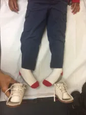
Below is a clinical demonstration of in-toeing with the knee being held in rectus position by the clinician, the tibia lies in an internally rotated position causing the foot to follow by in-toeing.

Often, children will sit on their feet, as shown below, which promotes an intoe gait


Standing Intoe with the knee straight (Right leg). This is true tibial torsion causing intoe (Below)
Below, Femoral anteversion, with the knee peaking in, which can cause intoe gait
