


Intraop arthroscopic pics of talar OCD surgery.


A series of intraop ankle arthroscopy pics of synovial chondromatosis with OCD talus and tibia and microfracture.
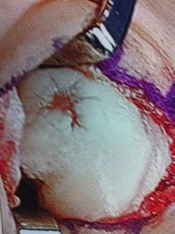
Pic of one of the nodules inside the ankle joint.


Pic of an OCD in the talar dome due to pressure from the nodule.

Pic after microfracture and debridement of above OCD.


Pic during microfracture of tibial OCD.


Pics of nodules removed during scope. A separate incision had to be made to remove the nodules due to the size.


Intraop arthroscopy pic of OCD afer debridement and during microfracture.


Intraop pictures of OCD of the talus after debridement and during microfracture.


OCD before and after microfracture.


Series of Pics of Microfracture of large OCD of the Talar Dome
Pic of OCD after preparation and debridement .

Measuring the size and depth of the OCD.


Action shot of microfracture of talus OCD.

Bleeding of OCD after microfracture.


First MPJ OCD (Below)

Osteochondral Autograft Transfer System (O.A.T.S.)


Chondrolysis of second MPJ arthritis before and after microfracture
There is damage to the articular surface cartilage which can occur either from an acute injury or more commonly after repetitive injury and/or wear and tear. If the bones at the joint in question are not aligned properly, then this mechanically adds to the wear that the joint receives with activity and increases the propensity of this occurring. Pictured below is linear damage to the articular surface of the second metatarsal head.

The following pictures are debridement of damaged cartilage that is abnormal in appearance and/or loose. This is removed completely to expose the subchondral (underneath cartilage) bone plate.


Drill holes are then placed through the bone plate to allow bleeding in the area, which will aide in healing of the cartilage surface with fibrocartilage, which is a kind of scarring of cartilage.

Series of Pics of OCD 2nd mpj (Below)
OCD with chondrolysis 2nd metatarsal head
Debridement and Curettage of loose and damaged cartilage


Microfracture/subchondral drilling of bone to stimulate fibrocartilage growth.
