pilon in French means "ceiling". In this injury it is referring to the ceiling of the ankle joint which is the articalar surgace of the tibia. Comminuted fracture from impact of the talus against the tibia articular surface (left). With high energy injuries such as a fall from a great height or a motor vehicle accident the communition could be very severe and involve both the tibia and fibula (right).

Post-surgical repair of the fracture with screws and plates used for fixation to peicemeal the shattered bone fragments back together. There are various types of fixation that a surgeon can use for this type of procedure. It depends on which devices work best to accomplish the goal of good reduction and re-alignment of the fragments as close to their original position as possible. pilon fractures are very complex injuries. This type of fracture involves the distal tibia and invades the ankle joint. They are usually associated with a fibular fracture. Common etiologies include car accidents, falls from heights and skiing accidents. These types of injuries usually are treated surgically due to the involvement of the joint surface, although some are either well aligned or so severe that they are treated non-surgically.
Series of pilon Fracture


pilon fractures have a 2 stage procedure for treatment. The first stage is usually an external fixator to hold the fracture out to length and in better reduction. That is kept on for 2-5 weeks until the soft tissue is in better condition. The ex fix is then removed and permanent ORIF is performed.
Below, Preop Displaced pilon Fracture


Below, After Ex Fix was placed to stabilize and temporarily reduce fracture


Below, Ex Fix is removed and ORIF performed. Reduction of fracture is complete.


pilon Fracture Before and after Ex Fix


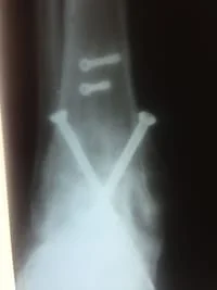
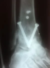
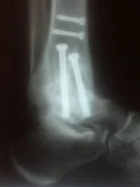
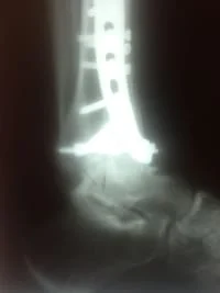
This a series of Xrays in a woman with Diabetes, poor vascularity and neuropathy. This injury was initially stabilized using an Ex Fix. After, definitive repair was performed using percutaneous plates and screws. This is a minimally invasive surgery that only requires small stab incisions to place the hardware. This significantly decreases the chances of complications.
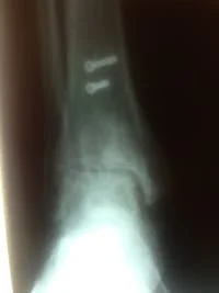
This is Preop xrays of the displaced pilon fracture


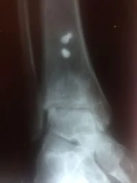
These are pictures of reduction with Ex Fix


These are intraoperative xrays showing the repair as well as small stab incisions used under fluoroscopic guidance


These are final intraop xrays showing excellent positioning and reduction of this devastating injury,


This is a pilon fracture with associated fibula fracture. Due to the high rate of complication, these are staged procedures. Either an ex fix or first reduction of fibula with our without internal fixation is performed. Once the soft tissue envelope is stabilized, a second procedure is performed to reduce the the tibia pilon fracture. In this case, an ORIF of the fibula was performed to reduce the fractures. Then a definitive ORIF of the tibia pilon fracture was performed.

These are pictures of a pilon fracture that was performed by a different surgeon. The anterior tibial plate was placed too distal and invaded the patient's joint. These injuries often cause post-traumatic arthritis regardless of the fixation. The plate was removed and the patient had some relief. Approximately 1 year later, the patient underwent a successful arthroscopic fusion.
S/P pilon with anterior plate too distal
After removal of the plate prior to arthroscopic fusion
After arthroscopic fusion