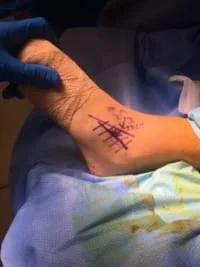
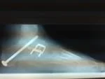
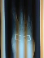
Intraop Pics of CN Bar resection in 11 year old gymnast. (Patient SH)
Incision Placement and the ICDN nerve mapped to avoid injury.

The Retinaculum and EDB muscle identified prior to dissection


The CN Bar is identified with the instrument and resected.


The CN Bar is completely resected.

The EDB muscle is visualized and transposed into the space created by the resection to decrease the chance of bone regrowth.


Pre and Postop X-rays of Above Patient

Set of Cn Bar Excision Pics Below
MIDDLE FACET COALITIONS
The following images are that of a 3D reconstruction of a CT scan that demonstrates the coalition of the middle facet of the subtalar joint.


3D reconstructions and CT scan of Middle Facet Subtalar Tarsal Coalition (Arrows Pointing to Coalition)


This image is a coronal plane view of a CT scan also showing a middle facet coalition.

Combination Coalition
The following is an example of a 12 y/o female with a combination coalition of the middle and posterior facets that has stopped her from doing atheletic activites because of pain. The top images are the lateral view (left) and oblique view (right) of the middle and posterior facet. The bottom left is the axial view of the middle facet.


The next set of images are CT scans in the transverse or axial view that demonstrate the irregularity of the facet and arthritic changes occuring at the area which cause the individual pain with activity. The bottom set of images are a sequence of the CT scan to show the irregularities from the coalition and its extent.


Pic of flatfoot treated with triple arthrodesis

Joints are realigned and in excellent positioning


