TENDONS
By: Robert H. Sheinberg, D.P.M., D.A.B.F.A.S., F.A.C.F.A.S.
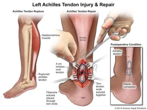
Tendons are firm, semi-flexible structures that connect muscles to bone allowing body parts to move.
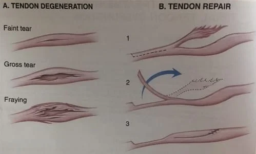
Injuries may be mild (tendinitis) caused by overuse of a body part. Moderate injuries are usually caused by stretching the tendon beyond its elastic limit causing a partial tear of the tendon. Complete disruption may be caused by severely stretching the entire tendon beyond its elastic limits or by a direct blow.
One of the worst injuries incurring the foot, ankle or leg region is a slow progressive weakening of the tendon that supports the arch. The arch will become progressively lower causing a flatfoot to appear.
Treatment of tendon injuries depends on making an early and correct diagnosis of the injured tendon and assessing the magnitude of the injury. Some injuries that are mild may require rest, ice and decrease in the intensity of the activity that caused the problem. Partial tendon tears usually require cast immobilization to prevent a complete tear. This is also often followed by physical therapy to allow return to sport or recreational activities. Complete tendon tears will almost always require surgery to repair the tendon.
Delaying the diagnosis and early treatment often aids in long-term failure. Despite late treatment for these injuries we can often restore complete function to the foot, ankle and leg, allowing a person or athlete to return to all their activities.
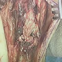
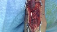
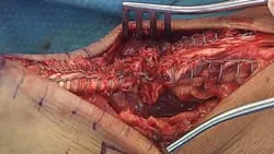
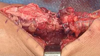
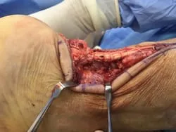
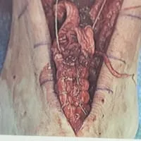
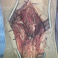
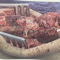
Intraop Pics of Ruptured 2,3 EDL Tendons after Boat Propeller Laceration
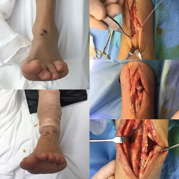
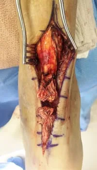
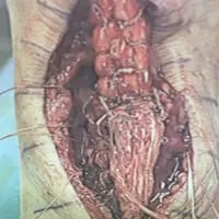
Series of Pics During Achilles Tendon Rupture Repair with Posterior Fasciotomy and FHL Muscle Transposition to Increase Vascularity to the Repair Site (Below)
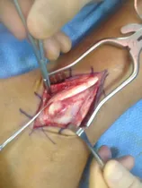
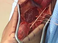
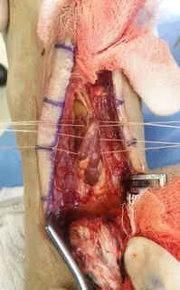
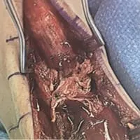
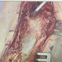
The rupture ends are at the top and bottom of the pic.
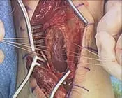
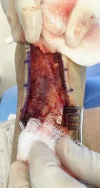
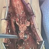
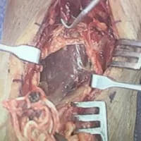
The window is made in the fascia between the Achilles and FHL muscle (muscle that moves the big toe down). We pass suture into the muscle and this can be seen in the fingers on the left and right of the pic.
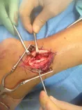
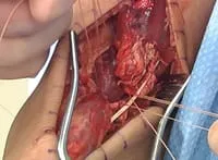
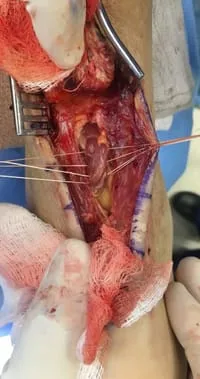
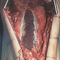
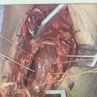
We have sutured the ends of the Achilles tear and these will be sutured together to repair the tear.

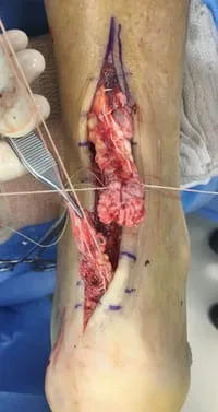
The tendon is tied together and the suture placed in the FHL muscle are tied around the tendon to tranpose the muscle to the tendon rapir to increase vascularization to the tendon and promote better healing.
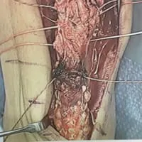
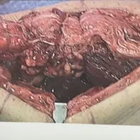
Intraop picture of Achilles tendon rupture. We have made a posterior fasciotomy to visualize the FDL muscle. We have 4 sutures in the center of the pic that are in the muscle belly. We tie these sutures on the pic of the tendon after the repair. This gives a better blood supply to the repair site.
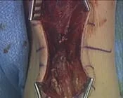
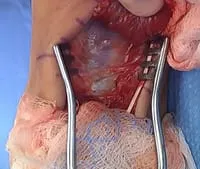
Pic of Rupture. The white tendon to the left is called the plantaris tendon. It doesn't give any tension to the Achilles or repair.
Windowing and Fasciotomy to Expose the FHL muscle
Pics of the Sutures through the muscle to transpose to the Repair site of the Achilles tear

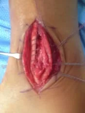
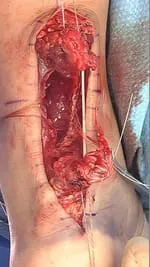
Krackow suture technique to lock the 2 ends of the repair site to decrease tension to the repair.
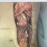
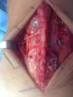
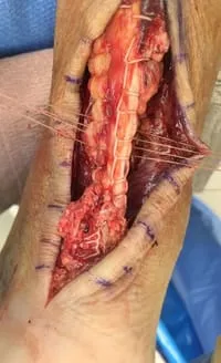
Repaired Achilles tendon with the sutures on the left and right holding the FHL muscle to transpose to the repair.

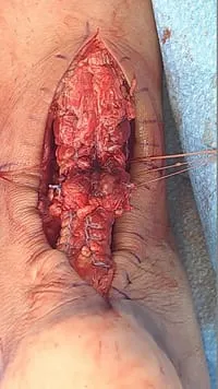
Pics of the complete repair and FHL muscle transposition to increase vascularity and healing to the repair

Intraop picture of Achilles tendon rupture. We have made a posterior fasciotomy to visualize the FDL muscle. We have 4 sutures in the center of the pic that are in the muscle belly. We tie these sutures on the pic of the tendon after the repair. This gives a better blood supply to the repair site.
Achilles tear
Pic of Fascia between Achilles and FHL muscle prior to fasciotomy to expose the FHL Muscle
Posterior Fasciotomy (Window) with FHL muscle exposed and the sutures seen at the right and left that will transpose the muscle to the repair.
Krackow suture technique on both sides of the Achilles tendon that locks the tendon on both sides of the Achilles to decrease tension at the repair site.

Repair of the Achilles. The sutures on the sides are attached to the FHL muscle prior to being tightened to transpose the muscle.
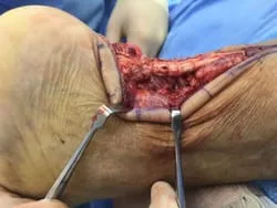
From the side, the FHL muscle can be seen transposed to the repair to increase vascularity and healing to the tendon.
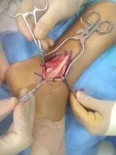
Series of Pics During Achilles Tendon Rupture Repair with Posterior Fasciotomy and FHL Muscle Transposition to Increase Vascularity to the Repair Site (Below)
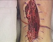
Torn Achilles Tendon
Identifying the Posterior Fascia that seperates the Achilles from the FHL muscle and compartment
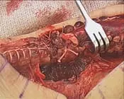
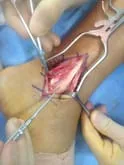
FHL Muscle is sutured across to bring better blood flow to repair site
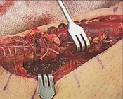
Krackow Suture of both Achilles rupture stumps
Rupture is Repaired and the FHL muscle can be seen as one with the Achilles to allow increased blood flow for healing.
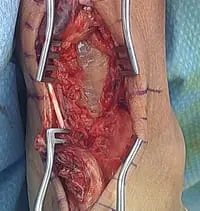
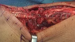
Intraop Pics of Achilles Repair with FHL Muscle Transposition
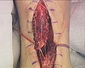
Rupture

Posterior Fasciotomy to expose FHL Muscle and Krackow Suture on both sides of the ruptured Achilles
The Muscle Transposed to the tendon after repair.
Intraop Pics of Achilles Repair with FHL Muscle Transposition
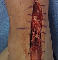
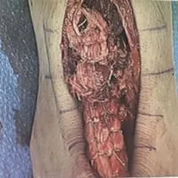
Pre and Post Repair of Achilles Rupture