In the pictures of MRI images below, the bone of the tibia shows high signal intensity (white within the black) that shows increased blood flow and swelling within the bone that indicates a stress reaction/fracture.


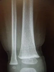
The x-ray image below demonstrates a fracture through the tibia that is in the process of healing. Most stress fractures do not show on x-ray until after they have started healing. It could take several weeks to see a stress fracture on plain x-rays.

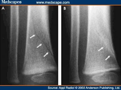
Subtle distal tibia spiral oblique fracture in young peds patient below.
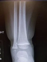
X-rays of nondisplaced toddler fracture
Subtle toddler fracture

Below is a stress fracture in a pediatric amateur athlete with fracture seen on X-ray, this is also referred to as "the dreaded black line".


Same fracture in CT scan seen 8 weeks later with bridging across the fracture and fracture callus.

