Types of Fractures:
- Diaphysis Fractures
- Metaphysis Fractures
- Stress Fractures
1. Diaphysis Fractures
The diaphysis is the long central portion of the tibia that attaches the proximal or rear portion of the bone to the distal or far part of the bone. Diaphyseal fractures can occur due to direct impact or a twisting or falling injury. If there is no displacement, these can be treated non-surgically with nonweightbearing cast and crutches. If the fracture is displaced, it may need to be treated in the operating room. It can be fixed with plates and screws, pins or a large intramedullary “IM” nail to place it in the correct position.
Below, Tibia and Fibula Fracture in a 16-year-old injured skiing. The patient presented to the office 8 days after injury with a displaced fracture. The first 2 films show the displacement in the fracture of the tibia. After an above knee cast is applied, the cast is wedged under fluoroscopy and new xrays show the near perfect reduction of the fracture. The white arrows show the area where the cast is wedged


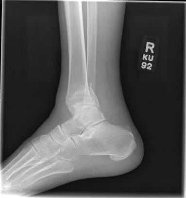
6 Months Post Injury and Back to Normal Activity


Wedging of cast to keep a tibia fracture well aligned. We wedge the cast on a side if there is malalignment early on in the fracture healing to realign the fracture


Immediate After Injury and 6 months post Injury back to normal activity


Xray of Tibia Fracture


Below is a post-surgical x-ray of a tibial fracture repair with screws and plate. This will stabilize and re-align the fracture fragment allowing appropriate healing.
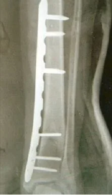
Pictures below show a 60-year-old woman who fell getting out of bed and broke the shaft of her tibia as has a distal fibular fracture. This was treated with an Intramedullary rod as well as a surgical plate and fixation with screws of the fibular fracture.


Displaced Tib-fib fracture that was treated with IM rod of the Tibia (below)


Below is a post-surgical x-ray of a tibial fracture repair with screws and plate. This will stabilize and re-align the fracture fragment allowing appropriate healing.
Pic of a 60-year-old woman who fell getting out of bed and broke the shaft of her tibia and has a distal fibular fracture. This was treated with an IM rod and plate and screw fixation of the fibular fracture.
2. Metaphysis Fractures
Fracture of the Tibial Metaphysis after traumatic injury

Preop CT Scan of Metaphysis fracture prior to repair below


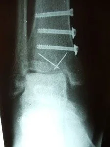
Below is a post-surgical x-ray of a fracture of the metaphysis after repair with screws and wires, one of which is a wire that was placed percutaneously (through the skin) and was then later removed.


Below is post-surgical film several weeks after repair after the percutaneous fixation pin was removed.
This a series of x-rays in a woman with diabetes, poor vascularity and neuropathy. This injury was initially stabilized using an Ex-Fix. After, the definitive repair was performed using percutaneous plates and screws. This is a minimally invasive surgery that only requires small stab incisions to place the hardware. This significantly decreases the chances of complications.
This is preop x-rays of the displaced pilon fracture

These are pictures of reduction with Ex-Fix
These are intraoperative x-rays showing the repair as well as small stab incisions used under the fluoroscopic guidance


These are final intraop x-rays showing excellent positioning and reduction of this devastating injury,


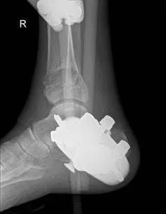
The following images are a case of a diabetic patient with a grossly displaced ankle fracture who initially thought she had suffered an ankle sprain. She became alarmed with the ankle joint started to displace with she was walking. She had not experienced a lot of pain because of the condition that diabetics can suffer from called neuropathy. The first set of images is pre-operative x-rays, which demonstrated the displacement.
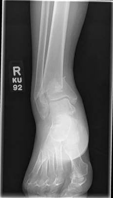

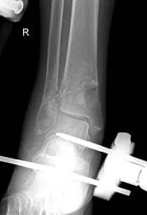
A staged provisional reduction was performed using an external fixator to control swelling and allow for a proper medical workup. X-rays are taken during surgery show re-alignment of the previously displaced fragments.


In the final stage of the repair (open repair with internal fixation), fluoroscopic images two weeks later indicated removal of external fixation and good reduction of the bone fragments with internal fixation using locked, minimally invasive, percutaneously placed plating and screws of the tibia and fibula. Anatomical alignment has been restored.


3. Stress Fractures
Problem:
- Microfractures that occur in any bone in the lower extremity or spine.
Causes:
- Overuse of the extremity may occur in an unconditioned athlete.
- Repetitive stress to a conditioned athlete without periods of rest.
- Malalignment of the lower extremity (excessively high arched foot or flat feet).
- Underlying bone demineralization (osteopenia, osteoporosis).
- Young females with menstrual irregularities.
Symptoms:
- Pinpoint tenderness to any bone in the extremity associated with localized swelling.
- Pain that gets progressively worse during an activity.
- As the problem progresses, pain can become severe causing an inability to weight bear or participate in any activity.
X-rays:
- Usually negative for the first 2-3 weeks, then as healing progresses x-rays may show bone callus formation.
MRIs:
- MRIs will usually show the earlier signs of stress fractures as the bone marrow starts to swell.
Treatment:
- Rest from the activity and cross training to continue overall fitness.
- Custom molded orthotic (shoe insert) to improve cushioning and realignment of the foot and lower extremity.
- Occasionally immobilization in a removable boot to allow the bone to completely rest and heal completely.
- Calcium and vitamin D supplementation.
- Bone stimulator if the fracture is not healing.
- Surgery in rare cases if the bone fails to heal.
In the pictures of MRI images below, the bone of the tibia shows high signal intensity (white within the black) that shows increase blood flow and swelling within the bone that indicates a stress reaction/fracture.


The x-ray image below demonstrates a fracture through the tibia that is in the process of healing. Most stress fractures do not show on x-ray until after they have started healing. It could take several weeks to see a stress fracture on plain x-rays.

This is a picture of a medial malleolar stress fracture prior to percutaneous screw fixation

Pics status post percutaneous repair with 2 screws


This is the same fracture seen on X-ray... the dreaded black line


The same fracture is seen 8 weeks later with bridging across the fracture and fracture callus


A stress fracture in former NFL football player

