Tibia Fractures in Children and Teenagers
Robert H. Sheinberg, D.P.M., D.A.B.F.A.S., F.A.C.F.A.S.
What is it?
- The tibia is the long bone between the knee and the ankle. It may be injured in toddlers, children, and adolescents.
How do they occur?
- Minor falls or twisting injuries in which the knee is locked or fixed in flexion and the foot externally rotates relative to the leg.
- Can occur in sports from direct or indirect trauma. Direct trauma would include a direct hit on the side of the leg by a helmet in football or possibly being kicked by another player during a sport such as a soccer.
- Common in motor vehicle accidents.
Where do they occur?
- Can occur in the upper one-third, middle one-third or lower one-third of the bone.
- Injuries to the central two-thirds of the bone heal much more slowly.
- They are often associated with fractures of the outer leg bone called the fibula.
- Fractures may be open in which the fractured bone tears through the skin and is open to the outside environment. This necessitates immediate surgical care to prevent infections and their devastating complications.
Signs and Symptoms:
- In a young child with a mild twist to the bone, a fracture may develop without any overt signs of swelling. Weight bearing may be difficult and the child will usually try to avoid putting pressure on the extremity.
- More significant injuries show acute signs of swelling and difficulty weight-bearing.
- It is important to make sure that the fracture has not opened through the skin.
- Deformity may be present in which the bone looks slightly crooked. It is a sign that immediate physician care is necessary.
- Numbness and tingling in the leg or foot can occur due to compartment pressures being elevated or a fracture that may be pressing on a nerve.
- Difficulty moving the foot up or down or toes in flexion or extension. If this elicits severe pain then immediate care is necessary to be sure that a compartment syndrome has not occurred.
- Other areas of injury must be sought out, which includes evaluation of the femur, knee and foot or ankle.
Immediate evaluation is necessary when the leg looks crooked or is the pain is severe. If the movement of the toes worsens symptoms, then a compartment syndrome may be developing, which requires attention immediately. If the bone is sticking out through the skin this is also an emergency that needs to be evaluated to prevent infections and other complications.
X-rays:
- These are always necessary not only to the tibia but to the ankle and knee to rule out other fractured bones.
CT Scan:
- May be necessary for severe injuries to evaluate the alignment not only of the fracture but of the ankle and knee if the fracture has gone through the joint.
- It is also utilized at times to check the healing of the fracture.
Compartment Pressures:
- Measuring the pressure in the compartments may be needed and done in an emergency setting to rule out an elevation of the compartment pressures, which could cause severe damage to the muscles, nerves, arteries, and veins.
Treatment:
90+% of all tibia fractures can be treated without surgery. Surgical emergencies include pending compartment syndrome or open fractures in which the bone is sticking through the skin.
1. Nondisplaced.
- If there is not an open fracture or compartment syndrome, then care can be planned without urgency.
- If the tibia has good alignment in all planes (side-side, forward-backward, inner versus outer turn) then immobilization in an above-the-knee cast may be necessary. Casting may be needed for 4-12 weeks depending on where the bone is fractured. If the tibia is fractured alone there is a tendency for the fracture to go into a virus (inner twist) alignment. After patients have an above-the-knee cast applied, weekly x-rays are taken for a month to evaluate healing of the fracture and to be sure the alignment of the bone is within normal limits. When the bone does not appear to be aligned perfectly, the cast is slightly pried open and small wedges of plastic are placed in the opening to realign the fracture during the healing process and to get the bone to heal straight.
2. Mildly Displaced.
- Mildly displaced fractures may require a gentle closed reduction that can usually be performed in the office without anesthesia. X-rays are taken and evaluated. Fluoroscopic images (live x-rays) may also be taken to gently realign the fracture to its normal position. Once the correct position is achieved, an above-the-knee cast is applied and weekly x-rays are taken to evaluate the healing of the tibia. Weekly x-rays are necessary because as swelling diminishes the cast may loosen, allowing the fracture to misalign. If this occurs and is caught early the fracture can be realigned into a more proper position to allow healing that is anatomic.
3. Moderately Displaced.
- Moderately displaced fractures may require a closed reduction under a general anesthetic. The child or teenager is put to sleep to avoid any pain and muscle spasm that may cause difficulty in reducing the fracture. While asleep, the fractured tibia is placed in its proper position. It usually can be held in this position with an above-the-knee cast. If it appears that the fracture is unstable, small pins may be placed through the skin to hold the alignment during the healing process. An above-the-knee cast is applied and the pins are removed simply in the office in 4-6 weeks or after sufficient fracture callus has developed to be sure that the alignment is going to stay. Rarely is an open reduction with plates or screws necessary to keep the alignment of the fracture.
4. Grossly Displaced.
- Grossly displaced fractures that are unstable will require surgery when closed reduction cannot be performed or if performed, instability still remains. Treatment may include closed reduction with or without percutaneous pinning and above-the-knee casting, elastic stable intramedullary nailing, minimally invasive plate osteosynthesis or rarely an external fixator. The treatment depends on the area of the bone that has been fractured and the experience of the surgeon performing these procedures. Close follow up monitoring is necessary to avoid complications with these fractures.
Complications:
- Malunion may occur when the tibia does not heal straight when looking at the leg from front to back or looking at the leg from side to side. The degree of malunion will dictate treatment. A slight malunion may not be problematic and if it occurs at a younger age has a chance to remodel into a more normal posture.
- The delayed union may occur when the tibia does not heal in a normal timeframe. If this occurs, continued immobilization may be necessary until signs of healing have taken place.
- Nonunions are more infrequent in tibia fractures in children and teenagers. If present, surgery may be necessary to help the bone unite.
- Missed compartment syndromes may occur if the pressure in the leg is elevated and has not been addressed early. Problems associated with compartment syndrome may include muscle contractures and a loss of use of certain muscle groups in the leg. Permanent numbness along an area that the nerve supplies sensation may also occur.
- Malalignment may put increased stress on the ankle, knee, and back, causing pain and possible premature wear and arthritis at a later time.
Tibia fractures, depending on the location, can take up to six months to heal. Close monitoring of the fracture with x-ray is important to be sure the alignment is maintained throughout the healing process. Although one closed reduction may be needed in most fractures, two or more closed reductions may be necessary for unstable fractures that want to continuously misalign. It is important to always evaluate the joint below (ankle) and above (knee) to be sure it has not been affected by the injury. An evaluation of the fibula is also important, as fibula fractures have to be taken into consideration in the healing of tibial fractures.
Below, Immediate After Injury and 6 months post Injury back to normal activity


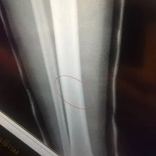
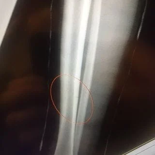
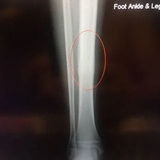
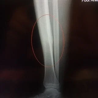
Below, Tibia and Fibula Fracture in a 16-year-old injured skiing. The patient presented to the office 8 days after injury with a displaced fracture. The first 2 films show the displacement in the fracture of the tibia. After an above knee cast is applied, the cast is wedged under fluoroscopy and new xrays show the near perfect reduction of the fracture. The white arrows show the area where the cast is wedged


6 Months post injury and back to normal activity.


Wedging of cast to keep a tibia fracture well aligned. We wedge the cast on a side if there is malalignment early on in the fracture healing to realign the fracture
Immediately after injury and 6 months post-injury back to the normal activity below.
Pre and Postop TIbia Shaft Fracture in 14 year old boy
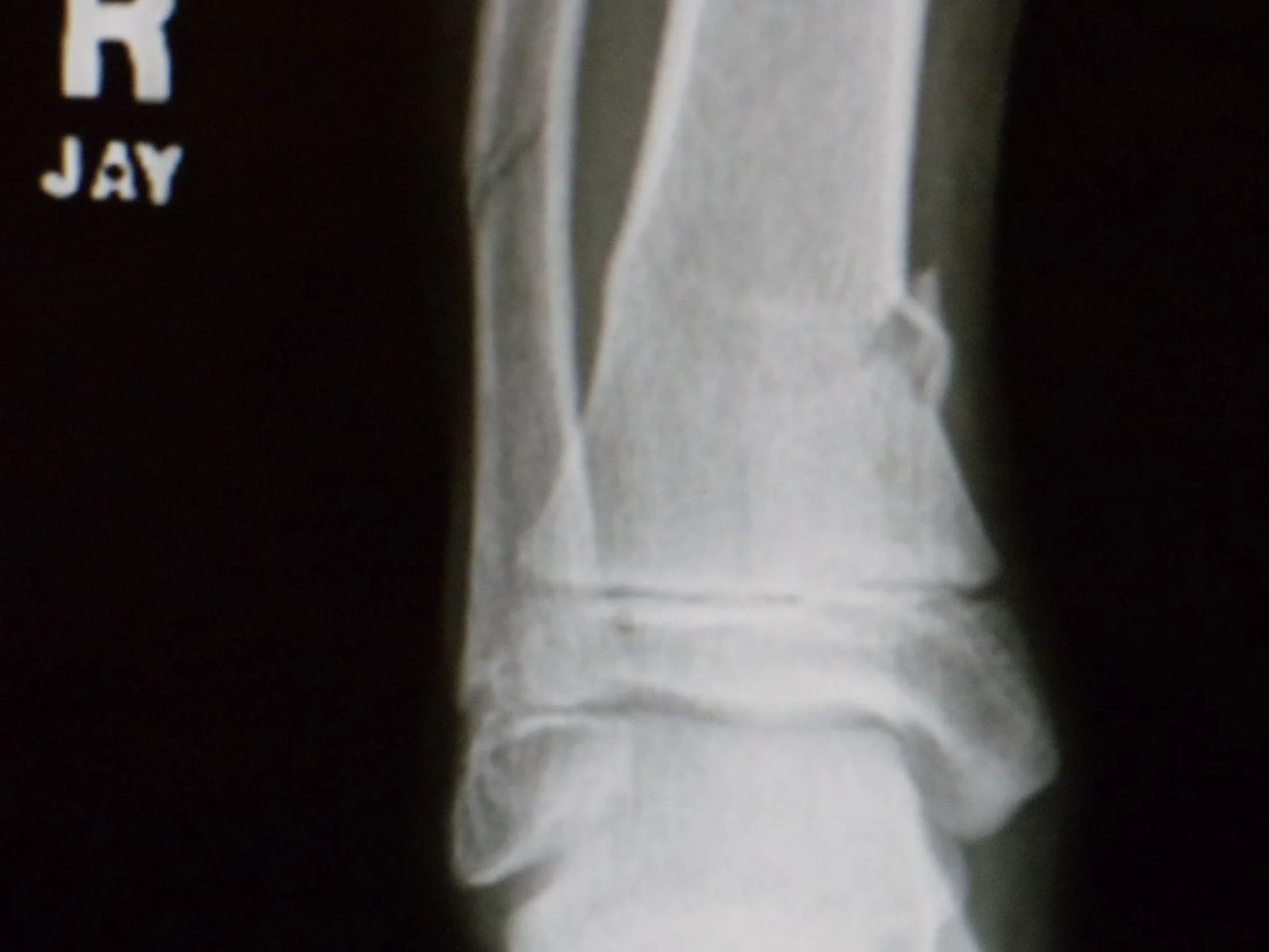
Fracture of the tibial metaphysis after traumatic injury